
Bruxism, Stress, and Adaptation
Many patients are diagnosed with bruxism or report that they grind or clench their teeth. But what exactly is bruxism? Over the years, the definition of this term has evolved considerably. Before exploring the topic in depth, let’s look at what a group of experts agreed upon during a consensus meeting held in 2017 (1). It is important to note that expert consensus does not automatically mean that the resulting conclusions are correct. It simply means that a group of recognised experts reached an agreement. Assuming that their definitions are true solely because they come from experts would be an appeal to authority fallacy ad verecundiam. With that in mind, let’s examine what was proposed.
For the first time, a distinction was made between awake bruxism and sleep bruxism. This distinction remains open to debate and will be analysed in greater depth in future articles. First, however, let’s review both definitions:
- Awake bruxism (AB) was defined as an activity of the masticatory muscles during wakefulness characterised by repetitive or sustained tooth contact and/or by bracing or thrusting of the mandible. It is not considered a movement disorder in otherwise healthy individuals.
- Sleep bruxism (SB) was defined as an activity of the masticatory muscles during sleep characterised by either rhythmic (phasic) or non-rhythmic (tonic) muscle activity. It is not considered either a movement disorder or a sleep disorder in otherwise healthy individuals.
The main novelty of these updated definitions is that bruxism is no longer classified as a disease or disorder in itself. Unless it is accompanied by signs or symptoms, it does not require treatment. To understand whether bruxism should or should not be considered a pathology, we first need to understand what unconscious or involuntary activity of the masticatory muscles actually does for the body.
Let’s look at several studies conducted in rodents in which different biomarkers were analysed. Unlike humans, rodents do not have an occlusion that allows them to grind their teeth together. Their dental anatomy does not permit tooth grinding in the same way as humans. Instead, their masticatory activity is centred on what their name implies: gnawing. To do so, they require an external object. This was precisely the basis of an interesting study published by Sato et al. (2008a) (2).
Initially, the researchers divided the animals (rats) into two groups: a control group and a stress group, in which stress was induced through physical restraint. Several biomarkers were analysed in both groups before and after the stress protocol, revealing statistically significant and clinically relevant findings:
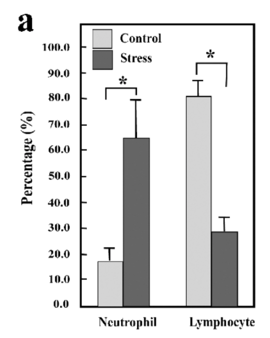
Blood samples were collected to analyse the ratio between T lymphocytes and neutrophils (both types of white blood cells involved in the neuroimmune system). Under non-stressed conditions, the ratio was approximately 80% T lymphocytes and 20% neutrophils. However, after six hours of physical restraint, this ratio progressively shifted to approximately 30% T lymphocytes and 70% neutrophils, indicating that stress induced marked changes in the neuroimmune system (Figure 1).
The most interesting finding came next: The researchers continued monitoring the animals after the stress period and allowed a subgroup of the stressed rats to gnaw on a wooden stick (simulating bruxism-like masticatory activity) during the following hours.
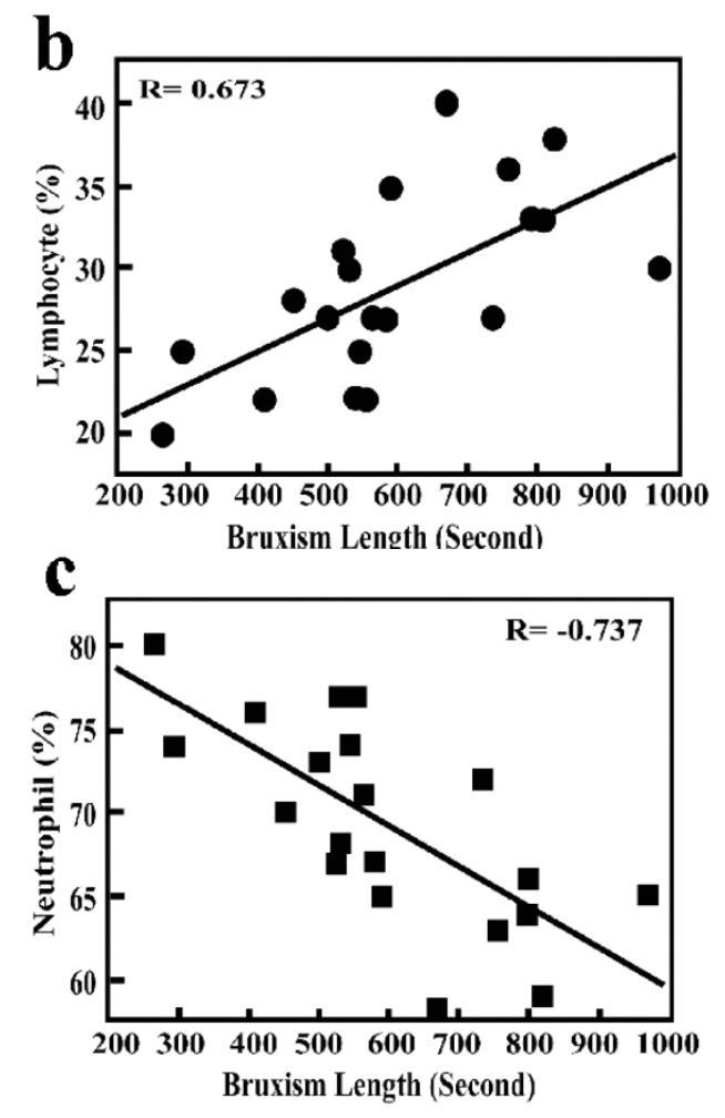
They observed that T lymphocyte levels, which had fallen during stress, gradually increased again, while neutrophil levels decreased, in proportion to the duration of the simulated bruxism activity. In other words, there was a correlation between the recovery of neuroimmune cell ratios and simulated bruxism during the hours following the stressful event (Figure 2).
Figure 1. Ratio of neutrophils to lymphocytes in the control group and the stress group.
Figure 2. T lymphocyte levels (b) and neutrophil levels (c) in relation to the duration of simulated bruxism following six hours of induced stress.
Stress induces changes in the neuroimmune system.
In addition, the size of the spleen and the thymus was measured. These glands are the primary sites for T lymphocyte maturation. Previous studies have shown that thymic atrophy is an indicator of neuroimmune system fatigue.
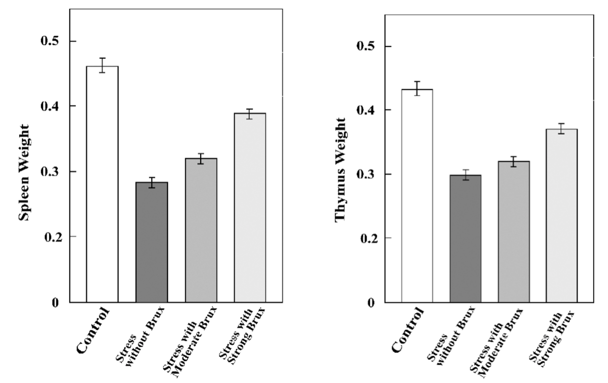
Within the stress group, the subjects were divided into three subgroups. The first group was allowed to gnaw on a wooden stick for an unlimited period (high simulated bruxism activity), the second group was allowed to gnaw for 30 minutes after the stressful event (moderate simulated bruxism activity), and the third stress subgroup was not allowed to perform any bruxism-like activity. The stress episodes were repeated until the gland sizes could be analysed.
The results showed that the greater the simulated bruxism activity, the more similar the size of the spleen and thymus was to that of the control group. In other words, simulated bruxism activity acts as a protective mechanism against atrophy of the spleen and thymus (Figure 3).
Figure 3. Size of the spleen and thymus according to the degree of simulated bruxism activity.
Therefore, so far we have observed what happens in the neuroimmune system during simulated bruxism activity at different levels. Apparently, this activity helps the body restore normal neuroimmune system parameters.
Let’s continue.
Perhaps we should investigate whether the stress we perceive is related to bruxism. Everything suggests that it is. At the hormonal level, when we are under stress, we secrete higher levels of glucocorticoids. In 2015, Karakoulaki et al. studied the relationship between perceived stress and cortisol levels in patients with and without bruxism. Perceived stress was measured using the Perceived Stress Scale (PSS), while cortisol levels were measured from saliva samples. Their findings showed that both perceived stress and cortisol levels were significantly higher in patients with bruxism than in the control group (3) (Figure 4).
Figure 4. Perceived stress and cortisol levels in patients with and without bruxism.
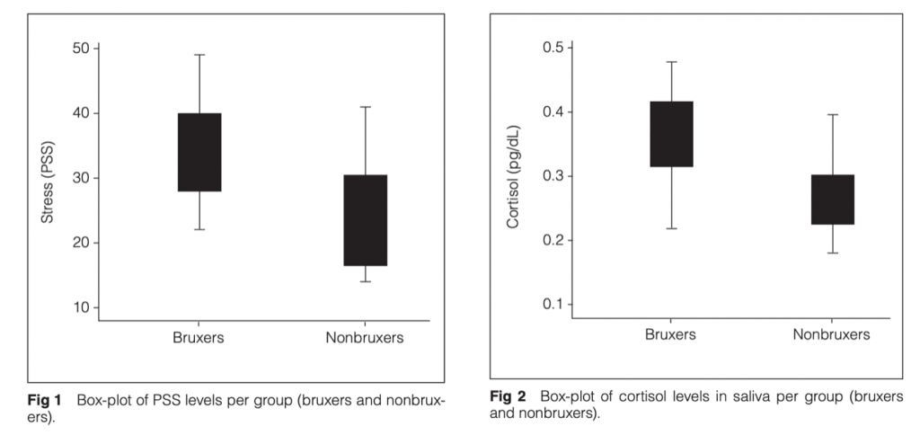
In other words, stress triggers a hormonal cascade, and it appears that the greater the perceived stress, the greater the bruxism activity, leading to changes in the immune system. We can therefore hypothesise that bruxism is a protective response by the body to the perception of stressful events.
And what happens in the brain? Let’s examine which brain areas become activated during stress and what happens if masticatory activity is simulated. This question was investigated by Sato et al. in 2008. They performed functional magnetic resonance imaging (fMRI) on subjects exposed to an unpleasant sound to induce acoustic stress, after which they were allowed to chew gum. During the stress exposure, several brain regions became activated. Notably, activity in the amygdala decreased significantly when the subjects chewed gum, whereas the brain regions involved in auditory processing remained active (Figure 5).
Figure 5. Amygdala activity in response to a stressful stimulus and to the same stimulus combined with masticatory activity.
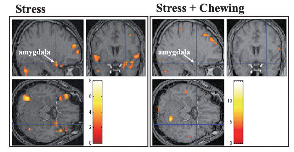
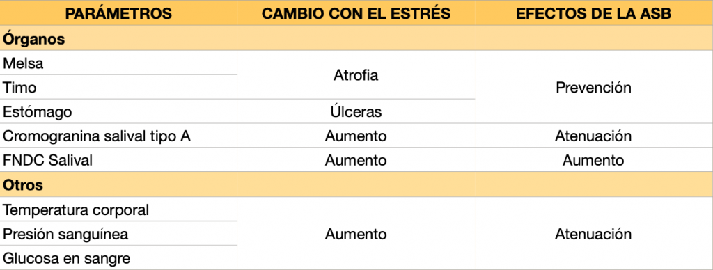
It appears that activity of the masticatory muscles may serve a protective function. Perhaps it is not a maladaptive response in the short term, provided that, with repeated exposure to the same stimulus, the body gradually responds with lower intensity—that is, provided that adaptation occurs through an allostatic process. Some of the adaptations observed by Sato and Slavicek (4) are summarised in Tables 1a–1c.
Table 1a. Summary of the effects of simulated bruxism activity on stress-induced changes in the brain.
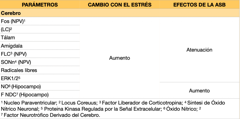
Table 1b. Summary of the effects of simulated bruxism activity on stress-induced changes in blood biomarkers.
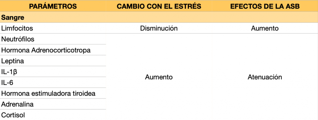
Table 1c. Summary of the effects of simulated bruxism activity on stress-induced changes in organs and other physiological parameters.
In response to stress, the body activates mechanisms whose purpose is to ensure survival and maintain well-being. These adaptive mechanisms were described by neuroanatomist Pieter Sterling, who introduced the concept of allostasis.
Allostasis means stability through change and refers to the processes by which living organisms adapt to current circumstances and prepare for similar situations in the future.
Figure 6. Diagram of the allostatic process.
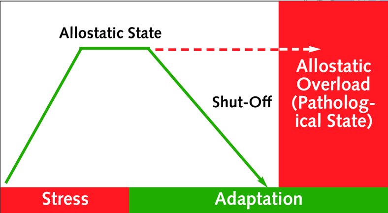
When stress is prolonged for too long or the body’s capacity to respond to stress is too low, it leads the body into a state of allostatic overload, or in other words, the adaptation that occurs will provide neither benefit nor harm to the individual in the future.
If it is stress-induced bruxism, the stress itself should be treated or the body should be helped to improve its physical adaptive capacities, either by redirecting the stress response towards muscles other than the masticatory muscles or by increasing masticatory capacity so that bruxism activity is not sufficient to cause pathology in the temporomandibular joint (TMJ).
CONCLUSIONS:
- Stress has effects at multiple levels, including the brain, immune system and organs. These effects can be beneficial if the body is able to adapt to them. If stress lasts too long or occurs so frequently that the body has not recovered from the previous stressful episode, it will not be able to adapt.
- Bruxism appears to be a response to cope with stress, which makes sense from a biological point of view, aiming to regulate the body through muscular activation, which in turn resembles the “fight” or “flight” stress responses.
- The role of bruxism appears to be more protective in nature, so it does not seem appropriate to focus on eliminating it without addressing the causes that trigger it.
- Bruxism may have consequences for the jaw and the temporomandibular joint (TMJ), but this is a topic that will be addressed in another Blog post.
______________________________________
BIBLIOGRAPHY:
1. Lobbezoo F, Ahlberg J, Raphael KG, Wetselaar P, Glaros AG, Kato T, Santiago V, Winocur E, De Laat A, De Leeuw R, Koyano K, Lavigne GJ, Svensson P, Manfredini D. International consensus on the assessment of bruxism: Report of a work in progress. J Oral Rehabil. 2018 Nov;45(11):837-844.
2. Sato S. et al. (2008) Bruxism and Stress Relief. In: Onozuka M., Yen CT. (eds) Novel Trends in Brain Science. Springer, Tokyo.
3. Karakoulaki S, Tortopidis D, Andreadis D, Koidis P. Relationship Between Sleep Bruxism and Stress Determined by Saliva Biomarkers. Int J Prosthodont. 2015 Sep-Oct;28(5):467-74.
4. Sato, S., Slavicek, R. The masticatory organ and stress management. J. Stomat. Occ. Med.1, 51–57 (2008).
RECOMMENDED READING:
Sapolsky, R. ¿Por que las cebras no tienen úlcera? (2016).
